
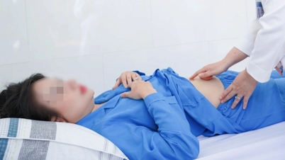
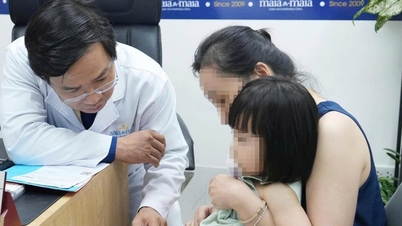
On October 1, Hue Central Hospital discharged patient HHMQ (6 years old, residing in A Luoi 2 commune, Hue city) who was diagnosed with right pulmonary arteriovenous fistula, a rare vascular malformation.
The patient was discharged from the hospital in stable health condition.
Previously, the child had a purple and fainting episode after eating cheese, was taken to a lower-level hospital for emergency treatment and then transferred to the Pediatric Center, Hue Central Hospital.
At the Department of Respiratory Pediatrics (Pediatric Center), the child was diagnosed with pneumonia and monitored for congenital heart disease. Upon admission, the child was thin, had purple lips, a blood oxygen level (SpO₂) of only 82%, and had a cough and mild difficulty breathing.
The doctor ordered blood tests, chest X-ray, echocardiogram and consultation with a Pediatric Cardiologist.
X-ray results showed pneumonia, transthoracic echocardiography only recorded mild left ventricular dilatation, heart function within normal limits.
These results were inconsistent with the patient's clinical condition, so the doctor suspected an abnormal aortopulmonary shunt and ordered a contrast echocardiogram.
The ultrasound results showed appropriate suspicion, so the patient continued to be assigned multi-slice computed tomography (CT 512 slices) of pulmonary blood vessels.
CT 512 results identified a pulmonary arteriovenous malformation (AVM) in the right lower lobe of the lung, about 6.5mm in diameter and 20mm in length, with a tortuous vascular bundle causing lobar pneumonia.
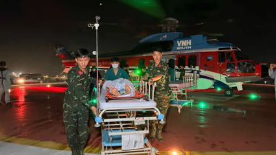
After intensive treatment of pneumonia with antibiotics, the patient was indicated for early intervention to close the fistula. On September 26, the intervention team including Stroke - Interventional, Pediatric Cardiology - Rheumatology, Anesthesia and Resuscitation doctors and a team of technicians performed the procedure.
Under endotracheal anesthesia, accessing via the femoral vein, the angiography team identified a high-flow pulmonary arteriovenous fistula, then used a specialized catheter to seal the fistula with a Coil combined with Onyx under a fluoroscopy (DSA) screen. After the intervention, angiography confirmed that the fistula was completely sealed, and blood oxygen saturation improved significantly, reaching SpO2 at 96%.
With stable vital signs, the patient was extubated and transferred to the Pediatric Cardiology - Rheumatology Department for monitoring. After three days, the child was alert, had no difficulty breathing, and was moving and eating normally.
Specialist II Doctor Nguyen Ngoc Minh Chau, Deputy Head of the Department of Pediatric Cardiology - Rheumatology, said that pulmonary arteriovenous fistula is a rare vascular malformation that often causes hypoxia, especially dangerous when the flow is large. The disease can be discovered by chance during examination or when complications appear.
Dangerous complications include stroke, brain abscess, transient cerebral ischemia, hemoptysis or pulmonary hemorrhage due to rupture of aneurysms.
In addition, the disease can also affect physical development, cause slow growth and heart failure. If not diagnosed and treated promptly, the disease can be life-threatening.
Currently, percutaneous intervention to close arteriovenous fistula is the main treatment method.
This is a minimally invasive technique, requires short anesthesia time, leaves no scars, has a quick recovery time and is less risky than open surgery.
The patient's mother shared that when she heard her child had a rare disease, the family was extremely worried.
During the treatment, the child was taken care of wholeheartedly by the doctors and nurses. Seeing the child healthy and playing with friends like now, the family is extremely happy./.
Source: https://www.vietnamplus.vn/hue-cuu-song-benh-nhi-bi-di-dang-mach-mau-hiem-gap-post1066224.vnp


![[Photo] Hanoi morning of October 1: Prolonged flooding, people wade to work](https://vphoto.vietnam.vn/thumb/1200x675/vietnam/resource/IMAGE/2025/10/1/189be28938e3493fa26b2938efa2059e)


![[Photo] President Luong Cuong receives President of the Cuban National Assembly Esteban Lazo Hernandez](https://vphoto.vietnam.vn/thumb/1200x675/vietnam/resource/IMAGE/2025/9/30/4d38932911c24f6ea1936252bd5427fa)
![[Photo] Panorama of the cable-stayed bridge, the final bottleneck of the Ben Luc-Long Thanh expressway](https://vphoto.vietnam.vn/thumb/1200x675/vietnam/resource/IMAGE/2025/9/30/391fdf21025541d6b2f092e49a17243f)


![[Infographics] An Giang economic picture in the period 2020 - 2025](https://vphoto.vietnam.vn/thumb/402x226/vietnam/resource/IMAGE/2025/10/1/093e29084648496c82a22536f5384c21)


Comment (0)