
For about a month now, Mr. NVT (49 years old, Hanoi ) has had chest pain behind the sternum when exerting himself, which subsides when resting. The patient has a history of smoking tobacco and cigarettes. Worried about his health, Mr. T. went to Medlatec General Hospital for a check-up.
Clinical examination and some tests, functional tests such as chest X-ray, electrocardiogram, cardiac enzyme test... the patient's indexes were all within normal limits. Because the symptoms and tests were not clear, the doctor ordered a coronary computed tomography angiography (CTA) to clarify the diagnosis.
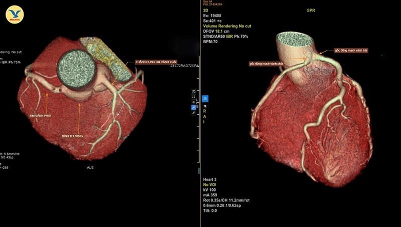
The scan results showed a rare congenital abnormality of the coronary arteries that supply the heart muscle: single coronary artery disease - type LII-B.
With this abnormality, the patient's left and right coronary arteries originate from a common root instead of originating from two separate locations, and are sandwiched between the pulmonary artery trunk and the aortic root, causing them to be congenitally stenotic. Specifically, in this case, the right coronary artery branch is narrowed by about 45% of its diameter.
According to specialist Dr. Tran Van Thu - Deputy Director of Medlatec Imaging Diagnostic System, a normal heart has two branches, the left and right coronary arteries, originating from two different locations in the coronary sinus of the aorta, from which blood will be carried to nourish the heart muscle.
When there is a single congenital coronary artery abnormality, there is only one common coronary artery originating from the coronary sinus and dividing into right and left coronary arteries to nourish the entire heart.
This is a rare congenital malformation, occurring in less than 0.05% of the population. Of these, type LII-B (Lipton classification) is a high-risk variant, where the right coronary artery originates from the left sinus of Valsalva and runs between the aorta and pulmonary artery trunk, easily causing myocardial ischemia and sudden death, especially during exertion.
Specialist Doctor I Tran Van Thu said that in diagnosing single coronary artery disease, coronary angiography with 128-slice CT scanner is the optimal diagnostic method, helping to accurately determine the starting position and path of the coronary artery; assess the degree of narrowing of the lumen and the spatial relationship with the large vessels; support treatment planning (medical or surgical).
If the path of the branch is favorable and the heart is still well nourished, no treatment is needed, just monitoring is needed. In case of symptoms or dangerous path of the vessel, consider surgery to reconstruct the origin or transposition.
The doctor added that most single coronary artery disease is discovered by chance, when the patient goes for a coronary CT scan due to suspected heart disease or for a general health check-up. Therefore, people should regularly have a periodic health check-up 1-2 times/year or when experiencing symptoms such as chest pain when exerting, shortness of breath, rapid fatigue, dizziness, etc., they should go to a medical facility for timely examination.
Source: https://nhandan.vn/dau-nguc-ban-co-the-mac-benh-tim-mach-chi-gap-o-duoi-005-dan-so-post913975.html

![[Photo] President Luong Cuong attends the 80th Anniversary of the Traditional Day of Vietnamese Lawyers](https://vphoto.vietnam.vn/thumb/1200x675/vietnam/resource/IMAGE/2025/10/09/1760026998213_ndo_br_1-jpg.webp)
![[Photo] General Secretary To Lam visits Kieng Sang Kindergarten and the classroom named after Uncle Ho](https://vphoto.vietnam.vn/thumb/1200x675/vietnam/resource/IMAGE/2025/10/09/1760023999336_vna-potal-tong-bi-thu-to-lam-tham-truong-mau-giao-kieng-sang-va-lop-hoc-mang-ten-bac-ho-8328675-277-jpg.webp)

![[Photo] Prime Minister Pham Minh Chinh chairs a meeting of the Government Standing Committee on overcoming the consequences of natural disasters after storm No. 11](https://vphoto.vietnam.vn/thumb/1200x675/vietnam/resource/IMAGE/2025/10/09/1759997894015_dsc-0591-jpg.webp)


Comment (0)