
Young, healthy people can still have strokes due to congenital heart defects.
In April 2024, Ms. M. (42 years old, Hai Duong ) suddenly suffered a stroke, leaving her with paralysis on the right side of her body. Despite receiving rehabilitation treatment and being examined at many medical facilities, she still could not find the cause of the disease. It was not until early August 2025, when she went to the hospital for examination, that the doctors were able to identify the "culprit" causing the stroke.
 |
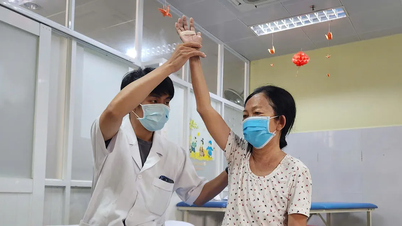
| The doctor is examining the patient. |
Dr. Nguyen Tuan Long, a cardiologist who treated the patient, said that the results of the in-depth examination showed that Ms. Mien had a patent foramen ovale, a small opening between the two atria, measuring about 3.5 mm. This was the cause of the previous stroke, through the mechanism of paradoxical embolism.
In normal people, small blood clots formed in the veins (especially in the lower limbs) will follow the blood to the right heart, then be pumped to the lungs to be retained and dissolved.
However, if the patent foramen ovale remains, in certain situations that suddenly increase pressure on the right side of the heart, such as coughing hard, sneezing or straining, a blood clot can "skip" through this hole to the left side of the heart. From there, it follows the bloodstream to the aorta and travels to the brain. If it gets stuck in the blood vessels of the brain, the clot will cause a blockage and lead to a stroke.
In Ms. Mien's case, if intervention to close the foramen ovale is not performed, the risk of stroke recurrence will be very high, seriously threatening her health and life.
Therefore, the doctors performed a minimally invasive intervention to close the foramen ovale. The team inserted a catheter from the femoral vein to the heart, located the foramen ovale, and used a 30 mm Occlutech PFO closure device to close the opening. The procedure lasted about 1 hour, after which Ms. Mien was conscious, her blood pressure was stable, and she was discharged after only 2 days of monitoring.
According to Dr. Long, Patent Foramen Ovale (PFO) is a fairly common congenital abnormality, accounting for about 24.2% of the population. During the fetal period, the lungs are not yet functioning, so blood will go from the right atrium to the left atrium through this natural "shortcut".
After birth, as the lungs begin to function, changes in heart pressure cause the foramen ovale to usually close on its own within the first few weeks or months of life. However, in about 25% of adults, the foramen ovale does not close completely, leaving a small opening.
Most PFOs do not cause symptoms and are considered benign. However, in some people, PFOs can lead to complications such as low blood oxygen causing shortness of breath, migraines, brain embolism or stroke. The risk of complications depends on the size of the hole, the amount of blood flowing through the hole and other risk factors.
PFO is the major contributing factor in approximately 50% of cases of ischemic stroke of unknown cause in young adults, according to the US National Library of Medicine (NIH).
According to the medical journal Frontiers in Neurology, patients with PFO in the high-risk group (with bypassed blood flow even at rest or a highly mobile atrial septum) have a stroke recurrence rate of up to 12.5% after 3 years, 3 times higher than the low-risk group (4.3%).
In people with stroke of unknown cause, this risk is up to 16.3%. In addition, the study also showed that large PFO (>4 mm) increased the risk of transient ischemic attack (TIA) 3.4 times, stroke 12 times and if there were ≥2 strokes, the risk increased up to 27 times.
Dr. Long recommends that patients under 60 years old who have had a stroke or transient ischemic attack without obvious cardiovascular risk factors should be screened for PFO.
Early detection and timely treatment will help prevent serious complications. Currently, patent foramen ovale occlusion is a minimally invasive and highly effective method, reducing the risk of stroke recurrence by up to 90% in appropriately indicated patients.
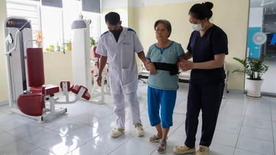
After the intervention, the patient can quickly recover movement and return to work, however, he/she needs to have a cardiovascular examination after 1 month and follow the doctor's instructions.
In addition, if warning signs such as chest pain, difficulty breathing, palpitations, severe headache, movement or speech disorders, unusual bleeding or bruising appear, go to the hospital immediately for timely treatment.
In addition, the doctor also emphasized the role of a healthy lifestyle in preventing cardiovascular complications. Patients should eat moderately, reduce salt, limit fatty foods and animal organs; increase green vegetables, fresh fruits and maintain drinking enough 1.5 liters of water per day.
Timely detection of stomach cancer when the tumor has not yet formed
Mrs. Huong, 69 years old, in Da Nang , went for a routine health check-up without any unusual symptoms. She had no underlying medical conditions, and no one in her family had gastrointestinal cancer.
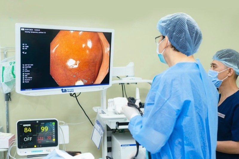
However, during the gastroscopy at the hospital, the doctors discovered a concave lesion in the body of the stomach at the lesser curvature, measuring only 0.8 cm, accompanied by changes in the surface structure and blood vessel system. Although the lesion was small and atypical, the doctor still decided to biopsy the tissue sample for further examination.
The biopsy results unexpectedly showed that Ms. Huong had early stage diffuse signet ring cell adenocarcinoma, a type of cancer that does not create a clear tumor but spreads silently in the mucosal layer, causing the stomach wall to thicken without any specific symptoms.
Subsequent CT scans showed no evidence of perigastric fat invasion, seroma, or other lesions.
Cancer cells are only in the mucosal layer, not yet invading blood vessels, nerves or neighboring organs. This means that the disease is detected extremely early and can be completely cured.
Doctors indicated intervention using endoscopic submucosal dissection (ESD), a modern, minimally invasive method that preserves the entire stomach.
During the procedure, the patient was given general anesthesia, the doctor inserted a flexible endoscope through the mouth to access the lesion, marked, cut and peeled the surrounding mucosa and removed the entire suspicious area for pathological anatomy. The entire procedure took about 30 minutes. After recovering, Ms. Huong did not feel any pain and was discharged after only one hour.
The post-procedure pathology results confirmed that the cancer cells were still confined to the mucosa, the cut surface was clean, and no additional treatment such as chemotherapy or radiotherapy was needed. Ms. Huong only needed to have regular check-ups for monitoring.
According to experts, detecting stomach cancer at an early stage is a key factor in increasing the survival rate to 90% after 5 years and the possibility of a complete cure.
However, in Vietnam, most stomach cancer patients are only diagnosed at an advanced or late stage, when the tumor has deeply invaded or metastasized, making treatment difficult and significantly reducing life expectancy.
Advanced cancer often requires total gastrectomy, lymph node dissection, combined with chemotherapy, radiotherapy or immunotherapy. The treatment process is complex, expensive and greatly affects the patient's quality of life. Therefore, regular screening is a vital factor.
Nowadays, with the support of advanced, high-resolution endoscopy systems, doctors can detect early lesions as small as a few millimeters, including flat lesions or lesions deep under the mucosa that were previously easily overlooked. Thanks to that, the rate of early cancer detection in Vietnam has improved significantly in recent years.
Experts recommend that people should proactively undergo regular gastric endoscopy after the age of 45, even from the age of 40 if there are risk factors, because gastric cancer tends to be younger. Even if there are no symptoms, early screening is still necessary to protect health and life.
Raising a 750g premature baby with brain hemorrhage
After 10 years of marriage and three failed IVF attempts, Ms. H. (31 years old, living in Ho Chi Minh City) finally became pregnant successfully through the fourth embryo transfer. Her pregnancy was closely monitored because of many risk factors for infertility, short cervix, threatened premature birth, and pre-eclampsia prevention from week 12 with aspirin.
At the 14th week of pregnancy, she had to have her cervix stitched. Severe morning sickness during the first three months combined with anxiety made her decide to quit her job to focus on her pregnancy because this was the child both families had been waiting for for a decade.
She thought her pregnancy was fine, but at week 25, when she was tested for gestational diabetes at Tam Anh General Hospital in Ho Chi Minh City, she was suddenly diagnosed with severe preeclampsia: blood pressure skyrocketed above 180/100 mmHg, high proteinuria, accompanied by leg swelling and headaches. Despite the doctor's efforts to prolong the pregnancy, at week 26, her blood pressure could not be controlled, and she was ordered to have an emergency cesarean section to save both mother and child.
The baby girl was born weighing only 750 grams, breathing weakly, and with severe respiratory failure. Right in the operating room, the neonatal team performed the “golden hour protocol” to support positive pressure ventilation and stabilize body temperature, then transferred the baby to the Neonatal Intensive Care Unit (NICU) for continued intensive resuscitation.
Dr. Nguyen Minh Thanh Giang, Neonatal Center, said the baby was diagnosed with severe respiratory failure due to immature lungs, requiring pulmonary surfactant injection. After that, the baby had a prolonged apnea, cyanosis, had to be intubated and put on a ventilator. After 24 hours, the baby's breathing improved, and he was switched to non-invasive ventilation.
Although the baby has overcome the first critical stage, his hemodynamic condition is still unstable due to his weak heart, not strong enough to pump blood to nourish the body.
Doctors continuously monitored blood pressure, used vasopressors, and adjusted acid-base balance to help the heart function effectively. Thanks to strict infection control, the baby only needed basic antibiotics and they were stopped early, not broad-spectrum antibiotics like most other extremely premature babies.
Along with respiratory and cardiovascular resuscitation, the baby was fed intravenously and given early milk. After 20 days, the baby was able to eat completely through the digestive tract.
However, by the fourth week, the ultrasound showed that the baby had a grade 2 cerebral hemorrhage, a condition in which blood had entered the ventricles but had not yet caused ventricular dilation. This is a common complication in extremely premature babies, because the blood vessels in the germinal area are very fragile and easily broken by sudden changes in blood pressure, oxygen, infection or stress.
The team strictly controlled the risk factors, kept the baby absolutely stable, avoided cyanosis, infection, hemodynamic instability. At the same time, blood transfusions and hemopoietic drugs were used to compensate for the lack of blood due to premature birth when the fetus had not yet gone through the important blood accumulation period in the last 3 months of pregnancy. After one month, the hematoma completely regressed, the baby's breathing was stable, no longer dependent on a ventilator, and practiced breathing with oxygen through a nasal tube.
In addition to medical efforts, mental factors are also given special attention. Dr. Cam Ngoc Phuong, Director of the Neonatal Center, said that the hospital applies the model of "Raising premature babies with family" to help parents overcome stress and confidently care for their children.
The mother was initially very worried and did not dare to hold her baby because “the baby was so small, I was afraid of hurting him”. The doctors provided psychological counseling and encouraged skin-to-skin contact to help reduce pain, stabilize the baby’s heart rate, improve immunity, increase mother-child bonding, and contribute significantly to the recovery process.
After more than three months in the hospital, the little girl now weighs 2.7 kg, equivalent to a full-term baby, can breastfeed and breathe on her own, no longer has a brain hemorrhage, is fully vaccinated and is eligible for discharge.
The family burst into happiness after the challenging journey. “The days of anxiety and fear are over. Finally, our family is complete when we bring our healthy child home,” the mother shared with tears in her eyes.
Source: https://baodautu.vn/tin-moi-y-te-ngay-119-nguoi-tre-khoe-van-co-the-dot-quy-vi-di-tat-tim-bam-sinh-d383622.html
![[Photo] Prime Minister Pham Minh Chinh chaired a meeting of the Steering Committee on the arrangement of public service units under ministries, branches and localities.](https://vphoto.vietnam.vn/thumb/1200x675/vietnam/resource/IMAGE/2025/10/06/1759767137532_dsc-8743-jpg.webp)

![[Photo] Prime Minister Pham Minh Chinh chairs a meeting of the Government Standing Committee to remove obstacles for projects.](https://vphoto.vietnam.vn/thumb/1200x675/vietnam/resource/IMAGE/2025/10/06/1759768638313_dsc-9023-jpg.webp)


Comment (0)