
The female patient had a 3x5 cm meningioma, which was operated on with a small 3 cm incision above the eyebrow, to crush and remove the entire tumor, for safety and aesthetics.
On June 13, Dr. Phan Van Dinh (Department of Neurosurgery, Neurology Center, Tam Anh General Hospital, Ho Chi Minh City) said that the medical team had just performed surgery on a meningioma in the skull base area of Ms. Duong Thi Hong (45 years old, Binh Thuan ), safely and aesthetically.
According to Ms. Hong, she had chronic sinusitis, often had dull headaches, went to massage and then it went away so she was subjective. Recently, her eyes were a bit blurry, she felt unwell so she went to Tam Anh General Hospital for a general check-up. The doctors ordered her to have a magnetic resonance imaging (MRI 3 Tesla), the results discovered a meningioma in the sella turcica (a part of the pituitary gland).
The tumor is 3x5 cm in size, located in the middle of the skull base, surrounded by many important components such as the anterior cerebral artery, optic nerve, pituitary gland... Doctor Van Dinh commented that skull base tumors are difficult to operate on, and if not careful, can leave many sequelae for the patient.
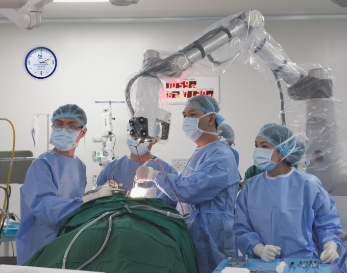
The patient is undergoing robotic surgery. Photo: Provided by the hospital
Previously, to operate on this type of tumor, the doctor had to make an incision in the skin and open the skull floor about 15 cm. Currently, thanks to the application of the keyhole technique, the doctor only needs to make a small incision about 3 cm right above the eyebrow to open the skull for surgery. This helps reduce the impact on the brain, limit the risk of brain retraction, brain contusion, and does not leave scars after surgery.
"This new surgical technique is more difficult. We use a combination of artificial intelligence brain surgery robots, microsurgery glasses, specialized Cusa machines to shrink tumors, and ultrasound to suck out tumors... This helps avoid the risk of rupturing blood vessels, nerves, or brain damage during surgery," said Dr. Van Dinh.
The surgery lasted 3 hours. After the surgery, the patient was awake, the tumor was gone, the nerve structure, the cerebral arteries... were preserved. The next day, the patient was able to walk and eat normally, his vision was restored and his headaches were gone.

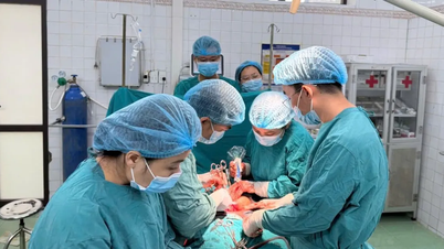
Doctor Van Dinh checks the patient's surgical wound. Photo: Provided by the hospital
Doctor Van Dinh said that most patients with meningioma often have symptoms of blurred vision and headache. Many people with blurred vision, double vision... only go to the doctor and treat eye symptoms. Only when the patient has blurred vision, paralysis of eye movements or severe vision loss, is the brain tumor discovered. At that time, the tumor has grown large, invading and affecting important brain parenchyma.
Through many surgeries for large meningiomas that have been left untreated for a long time, neurosurgeons warn that brain tumor symptoms are easily confused with other diseases. Therefore, when patients have symptoms such as unusual headaches, blurred vision, partial or total loss of vision, double vision, tinnitus, hearing loss, deafness, facial numbness, progressive weakness or paralysis, convulsions; memory loss, difficulty sleeping... they should go to the doctor for timely treatment. When the tumor proliferates, compressing the nerves and healthy brain tissue can easily endanger life.
Dawn
* Patient's name has been changed.
| In order to update the latest information on brain tumor surgery and cerebral hemorrhagic stroke using the Modus V Synaptive robot with artificial intelligence, the only one in Vietnam, Tam Anh General Hospital System organizes an online consultation week on VnExpress newspaper. The program takes place from June 8-14, readers can follow and ask questions here to get answers from doctors. |
Source link
![[Photo] General Secretary To Lam attends the ceremony to celebrate the 80th anniversary of the post and telecommunications sector and the 66th anniversary of the science and technology sector.](https://vphoto.vietnam.vn/thumb/1200x675/vietnam/resource/IMAGE/2025/9/29/8e86b39b8fe44121a2b14a031f4cef46)
![[Photo] Many streets in Hanoi were flooded due to the effects of storm Bualoi](https://vphoto.vietnam.vn/thumb/1200x675/vietnam/resource/IMAGE/2025/9/29/18b658aa0fa2495c927ade4bbe0096df)
![[Photo] National Assembly Chairman Tran Thanh Man chairs the 8th Conference of full-time National Assembly deputies](https://vphoto.vietnam.vn/thumb/1200x675/vietnam/resource/IMAGE/2025/9/29/2c21459bc38d44ffaacd679ab9a0477c)

![[Photo] General Secretary To Lam chairs the meeting of the Central Steering Committee on preventing and combating corruption, waste and negativity](https://vphoto.vietnam.vn/thumb/1200x675/vietnam/resource/IMAGE/2025/9/29/fb2a8712315d4213a16322588c57b975)


Comment (0)